February 8th, A proper face palm
Readers keeping track will notice that I skipped a post last Sunday. One reason could be because I was sightseeing during that weekend in London with Laura. A good reason withal, but perhaps not the real one. I suspect it has more to with the ruined experiment, the face palm of this week’s title. Let me explain.
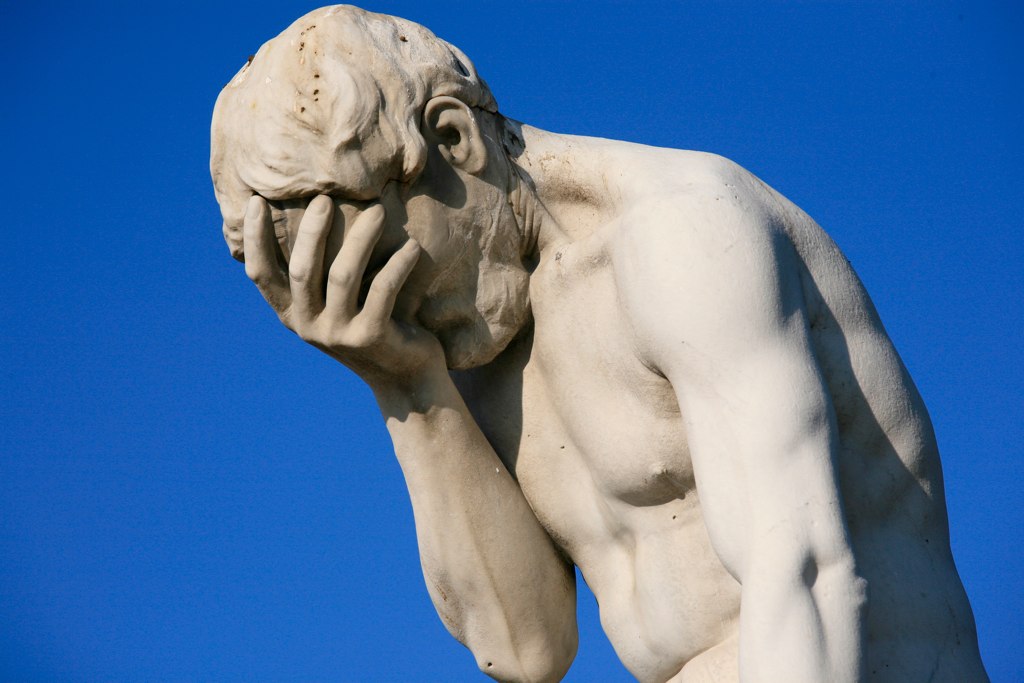
A classic facepalm, albeit for a more serious act than a blown experiment. This is Cain after killing Abel. See http://en.wikipedia.org/wiki/Facepalm
The experiment’s goal was to compare growth of coleoptile and mesocotyl. These are both embryonic organs, that is they are built by the special processes of embryogenesis and arrest as the seed the matures. Upon germination, they take up their major growth phase. All other shoot organs are built by the shoot meristem, which is why we use those special words. The coleoptile is a modified leaf that is rolled into a sealed tube, sheathing the first few leaves and meristem itself, presumably helping it go through soil on its way to the light. I believe the word coleoptile itself comes from a Greek word for a sword sheath. The coleoptile joins the stem at a node, below which is the mesocotyl, the first stem internode (below that is the primary root). Both mesocotyls and coleoptiles have been used in the classic stem-growth literature. It seemed reasonable to compare them. To accommodate the leaves, the coleoptile is hollow, which is an advantage in getting treatment compounds into the tissue, but doesn’t grow as fast as the mesocotyl in darkness, perhaps a disadvantage. Anyway, comparing them seemed like a good move.
The experimental plan was simple: cut coleoptile segments into one dish of buffer, mesocotyl into another, and then start the treatments. To improve the images, I transferred each segment from the buffer to the dry washer-plate before adding any treatment solution. The idea here was that rather than floating and sticking to each other, the segments would be on the glass substrate and separate. Once I had transferred the six segments, I put the washer-plate in the camera assembly, snapped an image, removed the plate, added the treatment solution, and finally put the plate on the shaker. The treatments were simple: control and 10 µM auxin. Only four washer-plates (two coleoptile, two mesocotyl). After 5 to 6 hours, I returned and removed the treatment solution by sucking it out with a pipette and imaged again dry segments for the final time point.
Well, when I came back for the final images, I discovered that, after taking the initial images, I had forgotten to add the auxin solution. Although he control washer-plates were both fine, the other two had segments dried to crisp. This was amazingly stupid. It was a small consolation, but only a small one, that this was the first such gaffe I have committed since August. Alas, I doubt it will be the last.
Nevertheless, it is a rare experiment from which nothing can be learnt. I was able to maneuver in the dark with more comfort, especially thanks to the wireless keyboard. Putting tape shapes on the washer-plates let me identify them individually with ease. The idea of imaging them ‘dry’ failed, because they were not dry, but instead were cloaked with a film of water, a shroud that made measurement worse (or would have had their been anything worth measuring!). But I did investigate the blurriness afterwards and re-focused the lens. I am not sure why the lens had come out of focus, but so indeed it had. This is sure to help. If it is not enough, then I will have to blot segments dry, which will be a minor nuisance.
I also learned that it is difficult to see where the mesocotyl starts even with infrared. In the light, the coleoptiles are a faintly yellow and there is a small ridge at the node. But in the infrared, the yellow vanishes and I could only once in a while see the ridge. Based on what I saw in the light in the post-experiment rubble, I am reasonably sure that in the first experiment I over estimated the length of the coleoptiles and consequently I think many of my mesocotyl segments in that experiment were taken from too far down steam (in the non-growing region). This makes an extra advantage for using coleoptiles, because at least their tip is unmistakable.
Next time, I am going to bring in a piece of black paper that I can put under the cutting area. Right now it is brightly reflective and looks just like the plant material. I want to see if the black paper will help contrast the node so I can see how long the coleoptile is and where the mesocotyl starts. Along these lines, I have a pen that paints in metallic silver and I want to see if I can see this ink in infrared. I tried a couple of regular writing pens and they are all invisible. It is helpful to write down which treatment is in which dish. I am going to try a coleoptile dose-response curve to auxin. I will use 3, 10, and 30 µM auxin. Many classic experiments use 10 µM. If the black paper helps me see the mesocotyl then next time I can do the same dose-response with mesocotyls. They don’t need to be compared in the same experiment. Stay tuned!